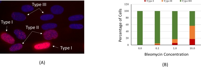
Fig 2.
(A) Category of cell nuclei stained with γ-H2AX antibody after bleomycin treatment. Nuclei with pan-nucleated staining are classified as Type I, Nuclei with distinct and countable foci are Type III, and nuclei with staining pattern in between are Type II. (B) Percentage of cells with different types of γ-H2AX staining after bleomycin treatment of 0.1, 1 and 10 mg/ml concentration. The percentage of Types I and II increased as the concentration of bleomycin increased.

