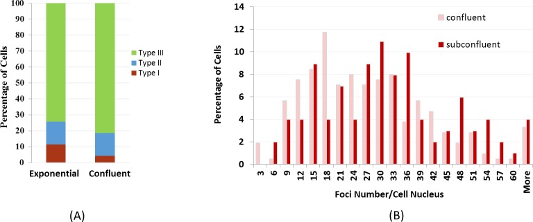
Fig 4. Percentage of nuclei with different types of γ-H2AX staining patterns in exponentially growing and confluent cells after bleomycin treatment of 1 mg/ml on the ground.
The percentage of Type I cells was significantly higher in exponentially growing cells. For bleomycin treated samples, the percentage of type I, II, and II are 11.5, 14.2, and 74.3 for exponential samples, and 4.3, 14.4, and 81.3 for confluent samples, respectively. (B) γ-H2AX foci number distribution in Type III nuclei in confluent and exponentially growing cells after bleomycin treatment showing differences in the lower quantiles of the two count distributions.

