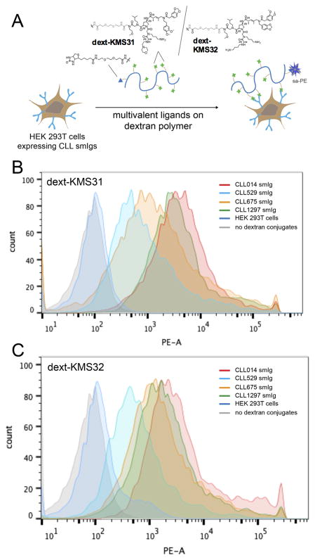
Figure 6. Flow cytometry analysis of ligand binding to cells displaying stereotyped CLL smIgs on HEK 293T cells.
(A) The cell binding assay with dextran conjugated multimeric ligands. CLL mAbs from subset 7p and other control CLL IgG were transiently expressed on the surface of HEK 293T cells. They were incubated with a biotinylated dextran polymer displaying 20–30 copies of the KMS31 or KMS32 ligands or controls followed by staining with phycoerythrin-conjugated streptavidin (sa-PE). (B) Histograms showing the binding of dext-KMS31 on the cells expressing stereotyped smIgs from subset 7P and controls, as analyzed by flow cytometry. (C) Same as B, but using dextran-conjugated KMS32. Reprinted with permission from ref. 25.

