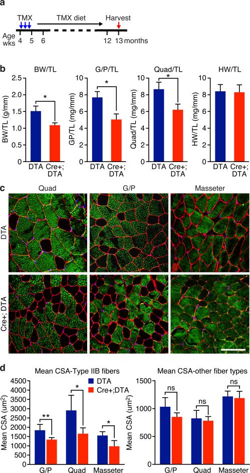
Figure 3. Ablation of the Tw2+ lineage causes type IIb myofiber atrophy.
(a) Schematic of TMX treatment. Tw2-CreERT2; R26-DTA/+ (Cre+;DTA) and R26-DTA/+ (DTA) mice on a mixed genetic background were injected with 1 mg TMX at 4 weeks of age on 3 alternating days. Mice were kept on TMX-containing diet until the time of analysis.
(b) Measurement of body weight (BW), heart weight (HW) and muscle mass normalized to tibia length (TL) of mice. Data are mean ± S.E.M; two sample t-test; *: P < 0.05. N =5 male mice for each genotype.
(c) Type IIb (green) and laminin (red) immunostaining of transverse sections of quad, G/P, and masseter muscles of Cre+;DTA and DTA mice. Scale bar: 100 um.
(d) Measurement of mean myofiber cross-sectional area (CSA) of type IIb fibers (left) and other fibers (right). Data are mean ± S.E.M; two sample t-test; *: P < 0.05. **: P < 0.01. ns: not significant. N =5 mice for each genotype. For each muscle, more than 300 fibers were measured and averaged.
Statistic source data for b,d are provided in Supplementary Table 3.

