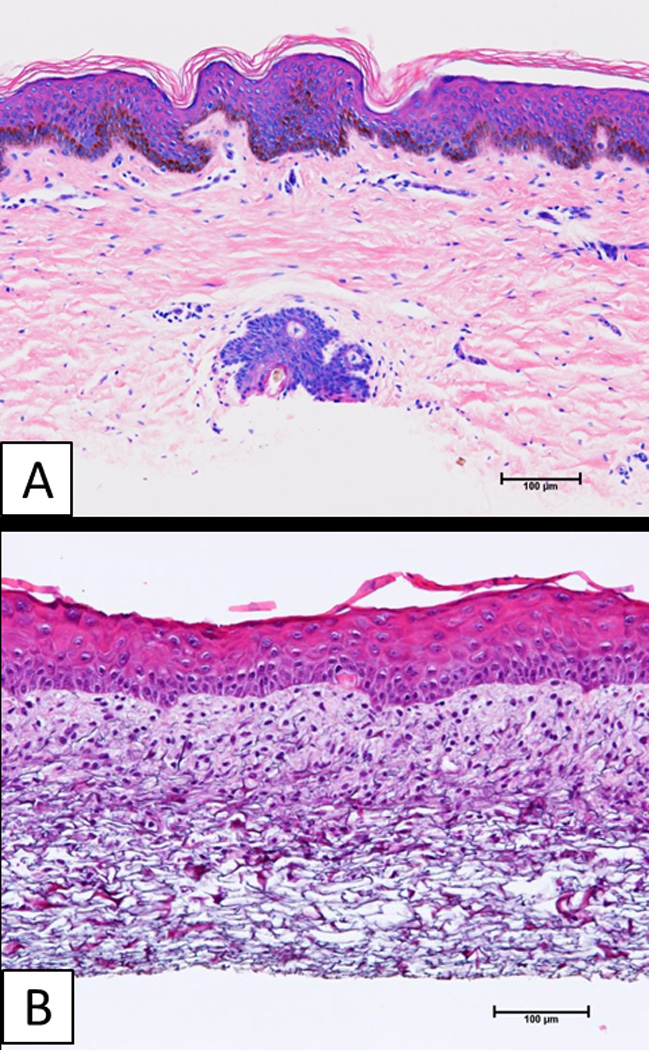
Figure 1.

Histologic anatomy of split-thickness skin autograft (AG), and engineered skin substitutes (ESS) prior to surgery. A) Split-thickness skin has a fully keratinized epidermis, and vascularized dermis with epidermal appendages. B) Engineered skin substitutes have partially keratinized epidermis, and a dermal substitute without a vascular network. Scale bar = 0.1 mm.
