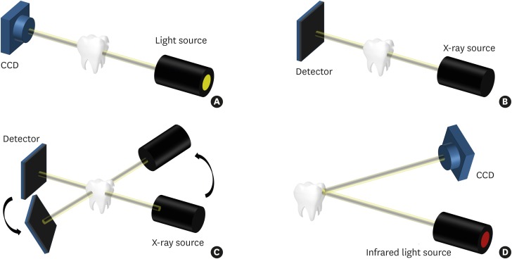
Figure 1.
The 4 imaging techniques used in the study. (A) Trans-illumination. A strong light is shone on one side of a tooth and the tooth image is acquired on the other side to identify cracks. (B) An intraoral X-ray is used to acquire a 2-dimensional image of a tooth using radiation. (C) CBCT is used to reconstruct a 3-dimensional image through image processing after obtaining radiographs from various angles. (D) OCT is used to acquire a cross-sectional image of a tooth using an infrared ray of 1,310 nm.
CBCT, cone-beam computed tomography; OCT, optical coherence tomography; CCD, charge-coupled detector.

