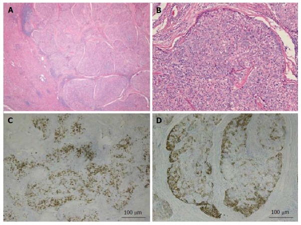
Figure 2.

Combined hepatocellular-cholangiocarcinoma with stem cell features, typical subtype. A: H and E, 4 × - tumor nests present on the right side with non-neoplastic liver on the left side; B: H and E, 10 × - peripheral small cells with hyperchromatic nuclei with mature appearing hepatocytes in the center; C: CK7, 4 × - scattered expression of CK7 by tumor cells; D: CK19, 4 × - patchy staining of the tumor and highlighting small tumor cells located at the periphery. Tumor was also positive for Hep-Par1 (not shown). H and E: Hematoxylin and eosin.
