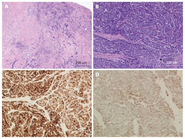
Figure 4.

Combined hepatocellular-cholangiocarcinoma with stem cell features, cholangiocellular subtype. A: H and E, 4 × - tumor cells present in tubular, anastomosing (antler-like) pattern; B: H and E, 10 × - small hyperchromatic tumor cells with high nuclear to cytoplasmic ratio present within dense fibrous stroma; C: CK7, 10 × - tumor is diffusely positive for CK7; D: CD56, 10 × - CD56 staining the cholangiolocellular component as well as the tumor cells at the periphery of the trabeculae. The tumor was diffusely positive for CK19 while negative for HepPar-1 and AFP (not shown). H and E: Hematoxylin and eosin.
