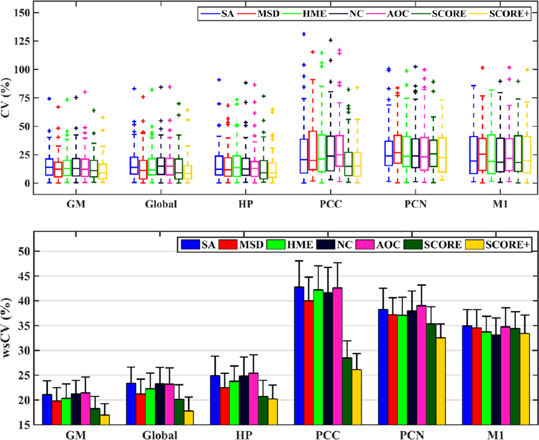
Fig. 5.
(TOP) Box plots of coefficients of variation (CV) for test-retest of CBF values in grey matter (GM), whole brain (global), hippocampus (HP), posterior cingulate cortex (PCC), precuneus (PCN), and primary motor cortex (M1) obtained using different algorithms. (BOTTOM) wsCV for different ROIs and for each method. The error bars show the standard errors in each case.

