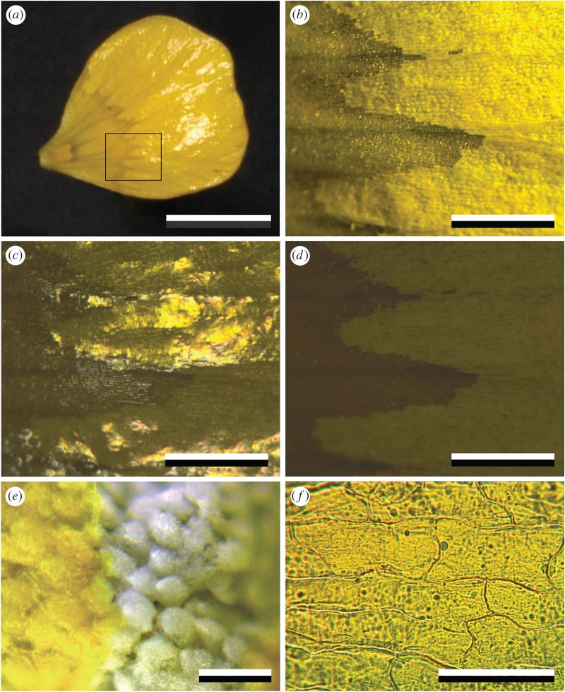
Figure 2.
Coloration of a glossy R. acris petal. (a) The glossiness is restricted to the main distal area of the upper (adaxial) side. (b) Close-up of the area indicated by the rectangle in (a), showing granular rows in the main petal area with oblique illumination. (c) The same area with normal epi-illumination. (d) The same area and illumination, but with crossed polarizer and analyser. (e) Close-up of a petal with disrupted upper epidermis, showing white starch cells. (f) Isolated upper epidermis observed in transmitted light. Scale bars: (a) 1 cm, (b–d) 1 mm, (e) 100 µm, (f) 50 µm.

