Abstract
[Purpose] To investigate the features of backward walking in stroke patients with hemiplegia by focusing on the joint movements and moments of the paretic side, walking speed, stride length, and cadence. [Subjects and Methods] Nine stroke patients performed forward walking and backward walking along a 5-m walkway. Walking speed and stride length were self-selected. Movements were measured using a three-dimensional motion analysis system and a force plate. One walking cycle of the paretic side was analyzed. [Results] Walking speed, stride length, and cadence were significantly lower in backward walking than in forward walking. Peak hip extension was significantly lower in backward walking and peak hip flexion moment, knee extension moment, and ankle dorsiflexion and plantar flexion moments were lower in backward walking. [Conclusion] Unlike forward walking, backward walking requires conscious hip joint extension. Conscious extension of the hip joint is hard for stroke patients with hemiplegia. Therefore, the range of hip joint movement declined in backward walking, and walking speed and stride length also declined. The peak ankle plantar flexion moment was significantly lower in backward walking than in forward walking, and it was hard to generate propulsion power in backward walking. These difficulties also affected the walking speed.
Key words: Stroke patients, Backward walking, Three-dimensional motion analysis
INTRODUCTION
In backward walking (BW) it is necessary to balance while moving backward, and this is difficult for elderly people and people with injury to the central nervous system, such as stroke patients. BW training and BW training on a treadmill are effective at improving measures of walking ability such as walking speed and stride length1, 2).
There are some reports of BW in healthy people3,4,5,6,7,8,9,10,11). Osugi et al.8) reported that walking speed, step length, and cadence were lower in BW than in forward walking (FW). Some studies3, 5,6,7, 10) have reported that joint movement patterns of the lower limbs are similar in BW and FW, whereas others4, 9, 11) have reported that they differ. Winter et al.5) reported that joint moments are similar for BW and FW. Opinion, therefore, varies widely.
We previously studied FW and BW in healthy university students12). Walking speed was arbitrary, and walking speed, stride length, cadence, peak hip joint extension, peak knee joint flexion, peak ankle joint plantar flexion, and peak hip and knee joint moments were significantly lower in BW than in FW. In addition, there was a difference in the timing of peak ankle joint moment, which showed that BW was not a simple reversal of FW.
In recent research, Fujisawa et al.9) reported that joint movement patterns differed between BW and FW. Additionally, Soda et al.11) reported that ankle joint power and work rate differed between BW and FW, and thus did not support the presence of similarities between FW and BW.
Although there have been reports of BW in healthy people, there are few reports of BW in stroke patients with hemiplegia. We believe that accurate analysis of the features of patients with paralysis will aid the development of more effective training programs and improve clinical medicine. Therefore, the purpose of this study was to investigate the features of BW in stroke patients with hemiplegia by focusing on the joint movements and moments of the paretic side, walking speed, stride length, and cadence.
SUBJECTS AND METHODS
Nine stroke patients with hemiplegia (eight male, one female; age: 68.9 ± 8.8 years; height: 162.7 ± 9.2 cm; weight: 63.4 ± 15.4 kg; five lower limb Brunnstrom stage IV, four lower limb Brunnstrom stage V; six brain infarction, three cerebral hemorrhage; average time since stroke: 6.4 years; two who using T-cane and brace on a daily basis) volunteered to participate. The requirements for participation were the ability to walk backward and forward more than 5 m without a brace or any other help, the usual ability of sight and understanding, and the absence of any serious disease or ailment in the waist or lower limbs. Participants were allowed to use a cane and two participants used a T-cane. All participants provided written informed consent to participate. This study was approved by the ethics committee of Hirosaki University Graduate School of Medicine (2014-076).
FW and BW were measured using a three-dimensional motion analysis system (Vicon Nexus; Vicon Motion System, Oxford, United Kingdom) with eight infrared cameras and a force plate (400 × 600 mm; AMTI, Watertown, MA, USA). Data were sampled at 100 Hz. Thirty-five infrared-reflective markers (14-mm diameter) were placed according to the Plug-in-Gait full-body model (Vicon). Data were analyzed using Polygon 4.
A 5-m walkway was prepared across the force plate and was marked using red tape placed on the floor. The red tape was placed in order to BW straightly without going off the walkway and to identify the finish line in BW. Participants were required to walk over the walkway at a self-selected pace while looking at the tape. The measurement order was FW followed by BW. Participants practiced both FW and BW several times. For safety, observers were present for every trial, but stood in a position that did not disturb the study.
Data were analyzed from the heel contact of the paretic side to the next heel contact of the same side in FW, and from the toe contact of the paretic side to the next toe contact of the same side in BW. Walking speed, the paretic side stride length, cadence, the percentage of the stride spent in stance, and joint angles and joint moments of the lower limb on the paretic side in the sagittal plane were quantified.
Statistical analysis of the data was performed using Statcel 3 (OMS Inc., Saitama, Japan). Statistical analysis included comparisons using the paired t-test for each variable measured during FW and BW. Two-tailed p values of less than 0.05 were considered statistically significant.
RESULTS
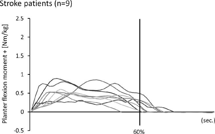
Temporo-spatial parameters are shown in Table 1. Walking speed and stride length were lower in BW than in FW. There was no significant difference in foot off between BW and FW. Peak joint angles are shown in Table 2. There was a significant difference in peak hip joint extension between BW and FW. Peak joint moments are shown in Table 3. There was a significant difference in peak hip joint flexion moment, knee joint extension moment, and ankle joint plantar flexion and dorsiflexion moment between BW and FW. Ankle joint moments in BW from all nine patients are shown in Fig. 1.
Table 1. Temporo-spatial parameters.
| FW | BW | |
|---|---|---|
| Walking speed (m/s) | 0.51 ± 0.15 | 0.21 ± 0.09** |
| Stride length (m) | 0.80 ± 0.14 | 0.36 ± 0.09** |
| Cadence (steps/min) | 75.86 ± 18.91 | 68.23 ± 16.70* |
| Foot off (%) | 60.52 ± 6.30 | 61.44 ± 8.93 |
Values are shown as mean ± SD. *p<0.05, **p<0.01
Table 2. Peak joint angles.
| FW | BW | ||
|---|---|---|---|
| Hip joint | Flexion (°) | 23.31 ± 7.61 | 23.31 ± 7.13 |
| Extension (°) | 8.58 ± 9.49 | −8.13 ± 9.29** | |
| Knee joint | Flexion (°) | 24.68 ± 8.06 | 22.06 ± 10.94 |
| Extension (°) | 1.92 ± 7.63 | 1.33 ± 8.14 | |
| Ankle joint | Dorsiflexion (°) | 14.50 ± 5.10 | 11.77 ± 3.51 |
| Plantar flexion (°) | 0.39 ± 8.13 | 2.16 ± 6.90 |
Values are shown as mean ± SD. **p<0.01
Table 3. Peak joint moments.
| FW | BW | ||
|---|---|---|---|
| Hip joint | Extension (Nm/kg) | 0.43 ± 0.16 | 0.56 ± 0.19 |
| Flexion (Nm/kg) | 0.46 ± 0.18 | 0.15 ± 0.09** | |
| Knee joint | Extension (Nm/kg) | 0.24 ± 0.12 | 0.06 ± 0.04** |
| Flexion (Nm/kg) | 0.37 ± 0.25 | 0.40 ± 0.25 | |
| Ankle joint | Plantar flexion (Nm/kg) | 1.09 ± 0.32 | 0.62 ± 0.19** |
| Dorsiflexion (Nm/kg) | 0.02 ± 0.01 | 0.02 ± 0.003* |
Values are shown as mean ± SD. *p<0.05, **p<0.01
Fig. 1.
Ankle joint moments in backward walking
DISCUSSION
The purpose of this study was to investigate the features of BW in stroke patients with hemiplegia by focusing on the joint movements and moments of the paretic side, walking speed, stride length, and cadence. Walking speed, stride length, cadence, peak hip joint extension, peak knee joint flexion, ankle joint plantar flexion movement range, and peak hip and knee joint moments were significantly lower in BW than in FW. These results are similar to the results of our previous study of healthy university students12).
Walking speed was arbitrary in this study as well as in our previous study12), and walking speed was lower in BW, which is an unfamiliar movement in daily life. The unfamiliar laboratory space may have also influenced walking speed and cadence, because patients took great care walking. Additionally, because participants were not required to maintain a set walking speed, this would have influenced cadence.
The peak hip joint extension angle was significantly lower in BW than in FW, and the hip joint movement range was narrower in BW. The decline in hip joint extension in BW was likely related to the shortening of stride length. We did not analyze the pelvis or the trunk in this study.
Kokubun et al.13) reported that swing of the paretic side lower limb was difficult for hemiplegia patients. To compensate for these swing difficulties, patients attempted to swing the paretic side leg by laterally bending the trunk to the non-paretic side and elevating the pelvis on the paretic side through hip joint abduction on the non-paretic side, or backward bending the trunk14). Consideration of the trunk and pelvis is required to fully understand BW in stroke patients with hemiplegia. Some patients tried to swing their leg backward by rotating their trunk and pelvis, and others tried to swing their leg backward by bending their trunk forward. In future studies, analysis of the trunk and pelvis will be required.
BW is a movement that involves conscious extension of the hip joint when stepping backward, which is different from FW. We believe that conscious extension of the hip joint is difficult for stroke patients with hemiplegia.
In our previous study12), we found no significant difference in the peak ankle joint flexion moment between BW and FW, and because there was a difference in the timing of the peak ankle joint flexion moment, it was not easy to generate propulsion power at the ankle joint in BW. In addition, Soda et al.11) reported that the propulsive force in BW must come from some factor other than the ankle. In the present study, peak ankle joint flexion moment was significantly lower in BW than in FW, and it was therefore difficult for participants to generate propulsion power. This affected the decline in walking speed.
Lee et al.10) reported that propulsion power and impact absorption occur primarily at the ankle joint, and the knee and hip joints do not contribute to propulsion power, and he emphasized the importance of the ankle joint in BW. In daily life, BW is not necessary over long distances, but is necessary for changing direction and avoiding falls. Therefore, BW does not require high walking speed. BW training and BW training on a treadmill improved walking speed and stride length1, 2). We believe that patients need to practice the push-off movement at the ankle joint to improve FW.
The ankle joint moment in BW varied widely in healthy university students. We believe that the risk of falls increases if the ankle plantar flexion moment increases and generates enough momentum to break one’s balance. Therefore, we think that there is a possibility that hemiplegia patients limit the ankle plantar flexion moment during BW.
In general, hip joint movement in the sagittal plane ranges from 30° of flexion to 10° of extension15). In the present study, hip joint movement in FW ranged from 23° of flexion to 9° of extension in stroke patients with hemiplegia, which is narrower than usual. Practicing BW without pelvis rotation and forward trunk bend may increase the range of hip joint movement.
One limitation of this study is that we did not measure trunk and pelvis movement. In addition, in contrast to FW, BW is not a familiar movement in daily life, and the strategy used for BW varies widely across individuals. Because there were only nine participants in this study, there are some features of BW that we were not able to uncover. In the future, we need to increase the number of participants and analyze trunk and pelvis movements and movement of the unaffected side as well as the affected side.
REFERENCES
- 1.Yang YR, Yen JG, Wang RY, et al. : Gait outcomes after additional backward walking training in patients with stroke: a randomized controlled trial. Clin Rehabil, 2005, 19: 264–273. [DOI] [PubMed] [Google Scholar]
- 2.Takami A, Wakayama S: Effects of partial body weight support while training acute stroke patients to walk backwards on a treadmill—a controlled clinical trial randomized allocation—. J Phys Ther Sci, 2010, 22: 177–187. [Google Scholar]
- 3.Thorstensson A: How is the normal locomotor program modified to produce backward walking? Exp Brain Res, 1986, 61: 664–668. [DOI] [PubMed] [Google Scholar]
- 4.Vilensky JA, Gankiewicz E, Gehlsen G: A kinematic comparison of backward and forward walking in human. J Hum Mov Stud, 1987, 13: 29–50. [Google Scholar]
- 5.Winter DA, Pluck N, Yang JF: Backward walking: a simple reversal of forward walking? J Mot Behav, 1989, 21: 291–305. [DOI] [PubMed] [Google Scholar]
- 6.Grasso R, Bianchi L, Lacquaniti F: Motor patterns for human gait: backward versus forward locomotion. J Neurophysiol, 1998, 80: 1868–1885. [DOI] [PubMed] [Google Scholar]
- 7.Kitayuguchi J, Minami S: Biomechanics of backward walking. J Phys Ther, 2003, 20: 551–556(in Japanese). [Google Scholar]
- 8.Osugi H, Miwa K, Shigemori K: Characteristics of backward walking of healthy adult. Rigakuryoho Kagaku, 2007, 22: 199–203(in Japanese). [Google Scholar]
- 9.Fujisawa H, Yoshida T, Onobe J, et al. : A study on speed control during backward walking for young adults. J Jpn Phys Ther Assoc, 2010, 37: 17–21(in Japanese). [Google Scholar]
- 10.Lee M, Kim J, Son J, et al. : Kinematic and kinetic analysis during forward and backward walking. Gait Posture, 2013, 38: 674–678. [DOI] [PubMed] [Google Scholar]
- 11.Soda N, Ueki T, Aoki T: Three-dimensional motion analysis of the ankle during backward walking. J Phys Ther Sci, 2013, 25: 747–749. [DOI] [PMC free article] [PubMed] [Google Scholar]
- 12.Makino M, Takami A: Comparison of forward walking and backward walking in healthy university students. J. Health Sci.Res. 2015, 5: 33–41. [Google Scholar]
- 13.Kokubun T, Tagughi T, Hoshi F: Body mechanics and physical therapy on walking in stroke hemiplegia patients. J Phys Ther, 2015, 32: 46–54(in Japanese). [Google Scholar]
- 14.Kerrigan DC, Frates EP, Rogan S, et al. : Hip hiking and circumduction: quantitative definitions. Am J Phys Med Rehabil, 2000, 79: 247–252. [DOI] [PubMed] [Google Scholar]
- 15.Perry J, Burnfield JM: Gait analysis normal and pathological functional, 2nd ed. New Jersey: SLACK, 2010, pp 103–119. [Google Scholar]


