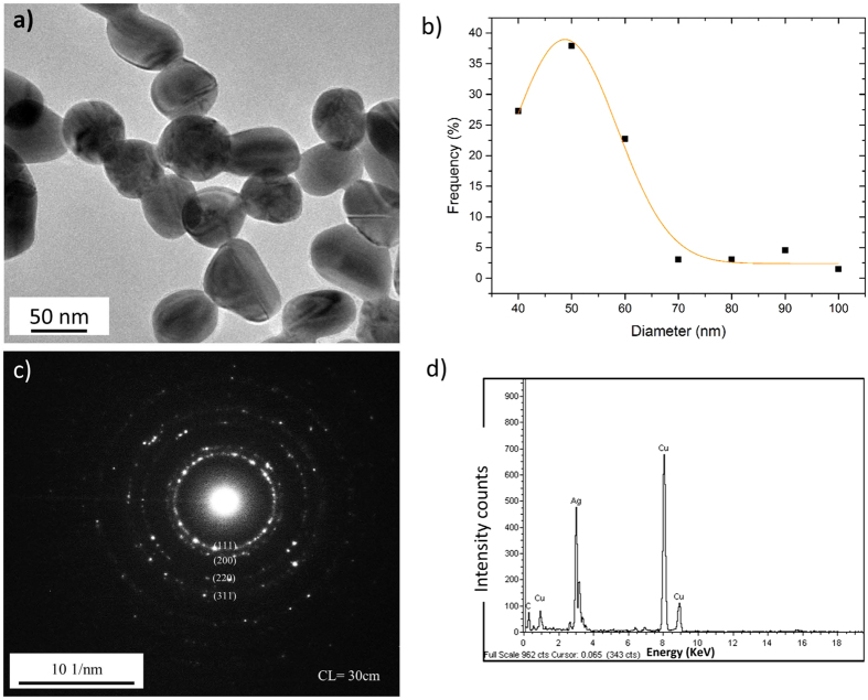
Figure 1. Characterization of the synthesised AgNPs.
(a) TEM image of synthesised AgNPs and (b) their size distribution measured through TEM. (c) Indexed selected area electron diffraction (SAED) patterns of AgNPs incubated in DI water, revealing characteristic lattice spacings of 0.236, 0.204 and 0.145 nm, corresponding to the (111), (200) and (220) planes of metallic silver. The selective area aperture size used was 100 nm in diameter. (d) EDX spectra of AgNPs. Data shown are representative of analysis of 200 particles.

