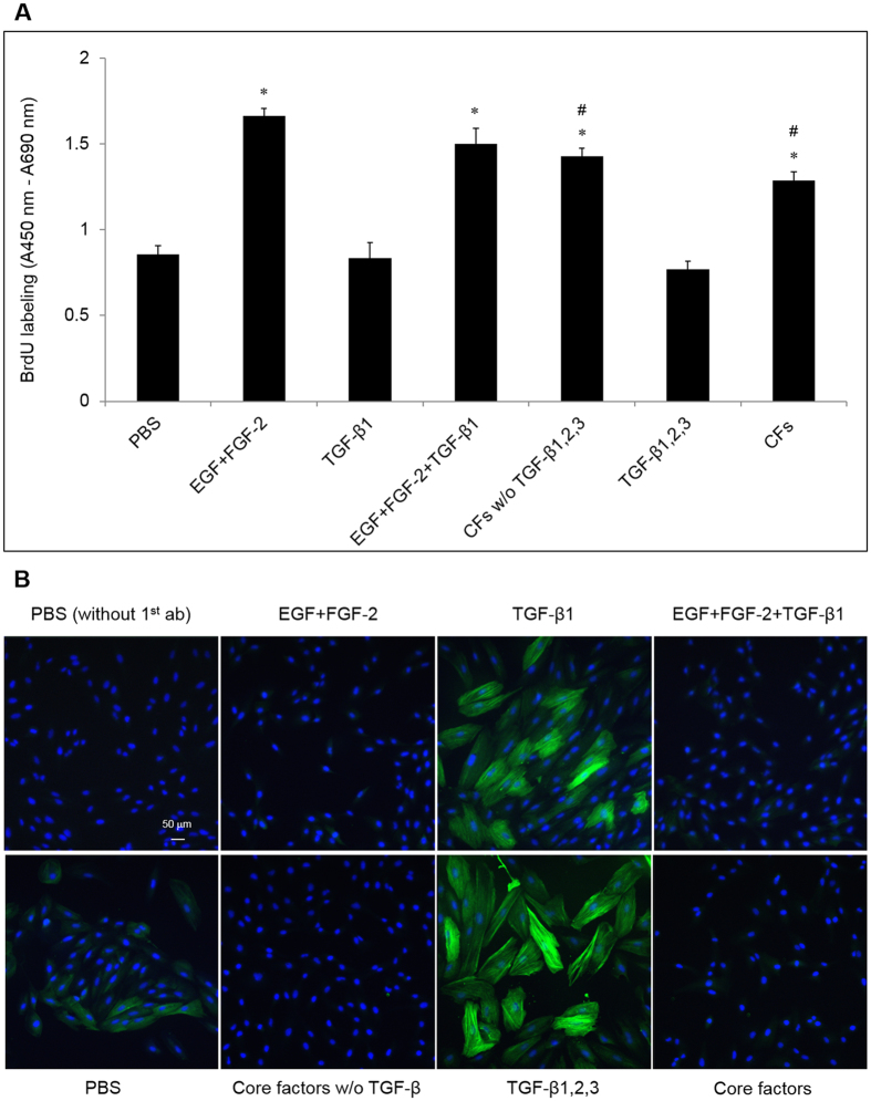
Figure 2. Proliferation and EMT affected by EGF + FGF-2 + TGF-β1 and the “core factors” (CFs).
(A) BrdU labeling (A, n = 3, *indicates p < 0.05 compared to the PBS control and # indicates p < 0.05 compared to EGF + FGF-2) and immunostaining α-SMA (B, nuclear counterstaining with Hoechst 33342, scale bar = 50 μm) of ARPE-19 cells seeded at 1 × 10⁴/cm2 in DMEM/F12/10% FBS for 24 h and then treated with PBS or EGF (10 ng/ml) + FGF-2 (20 ng/ml) (EGF + FGF-2), TGF-β1, β2, β3 (each at 10 ng/ml), or CFs (see Table 1) for 48 h.

