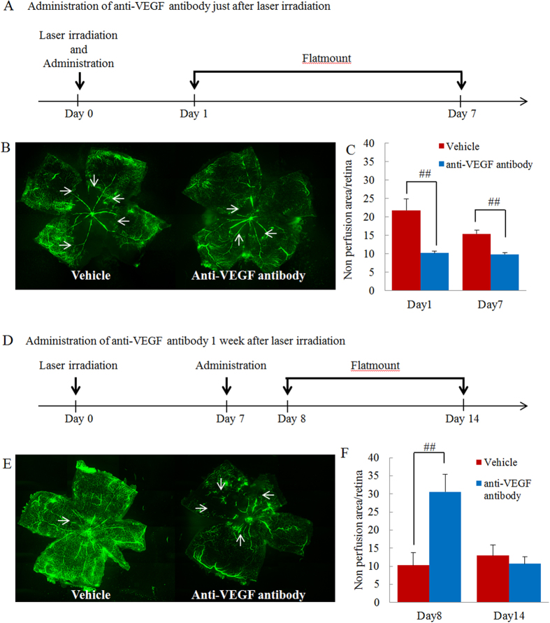
Figure 8. Anti-VEGF antibody prevented progression of retinal nonperfusion when administered in the early phase after occlusion but aggravated nonperfusion when administered in the late phase.
(A) Protocol of early phase administration. Anti-VEGF antibody was intravitreally administered immediately after occlusion. Sampling was performed 1 and 7 days after administration. (B) Representative images of flat-mounted retinas (vehicle and anti-VEGF antibody treated groups). (C) Illustration of quantitative retinal nonperfusion area data. Nonperfused areas were reduced 1 and 7 days after occlusion. Data are expressed as means ± S.E.M. (n = 4–8). ##P < 0.01 (Student’s t-test). (D) Protocol of late phase administration. Anti-VEGF antibody was administrated intravitreally 7 days after occlusion. Sampling was performed 1 and 7 days after administration. (E) Representative images of flat-mounted retinas (vehicle and anti-VEGF antibody treated groups). (F) Quantitative illustration of retinal nonperfusion area data. The nonperfusion area was increased by anti-VEGF antibody 1 day after administration but was unchanged 7 days after administration. Data are expressed as means ± S.E.M. (n = 4–5). ##P < 0.01 (Student’s t-test).

