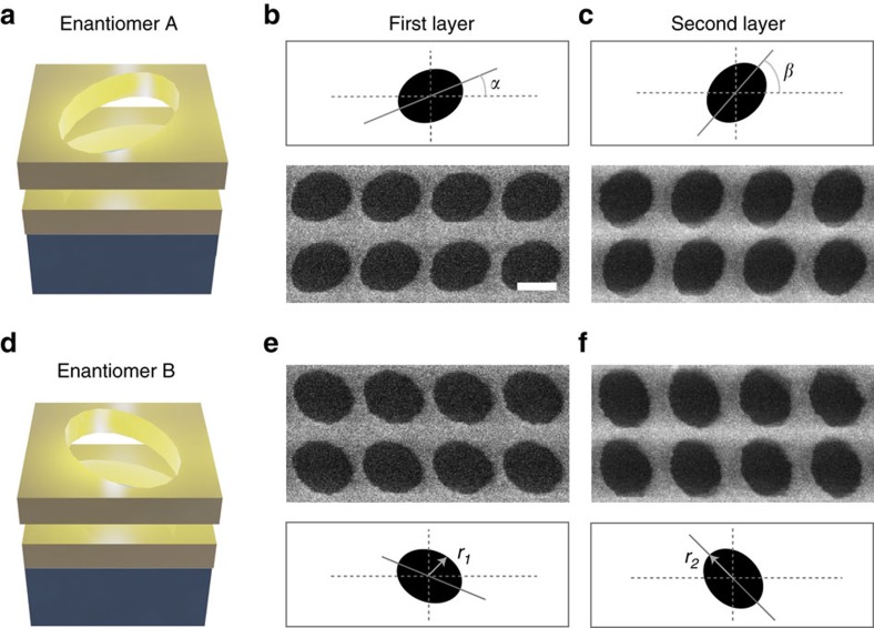
Figure 1. Schematic and micrographs of chiral metamaterial enantiomers.
(a) A single unit cell of the metamaterial array. The nanoengineered material is composed of two silver films separated by 45 nm of a dielectric spacer (n=1.45) and perforated with two ellipses, where the major axes of the ellipses are skewed by 22.5°. (b) The SEM image depicts the first layer of the structure and is followed by an image of the (c) upper layer placed on top of the first. The scale bar in the image is 200 nm and the overall pitch of the unit cell is 350 nm. The characteristic parameters of these ellipses are r1=115 nm, r2= 150 nm and α=22.5°, β=45° from the coordinate axis. The second unit cell on the lower half of the figure is then created by aligning the ellipses such that the unit cells are mirror images of each other known as enantiomers. (d) Schematics and SEM images of the (e) lower and (f) upper layers of enantiomer B are provided.

