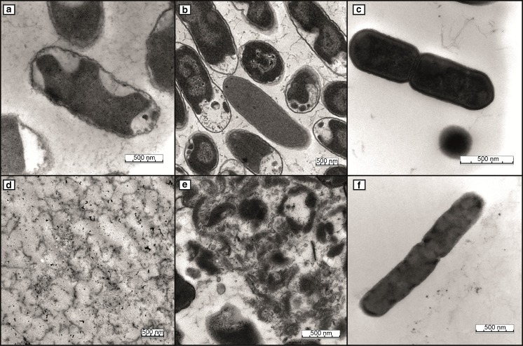
Fig. 4.
Transmission electron microscopy micrographs after the different extraction protocols with [BMPyr+][Ntf2 -]. Micrographs of Salmonella Typhimurium (a), E. coli (b), and Listeria monocytogenes (c) controls. For the final extraction protocol, 1 min and 150 °C were selected for all three species (d Salmonella Typhimurium, e E. coli, and f L. monocytogenes). Bars 500 nm

