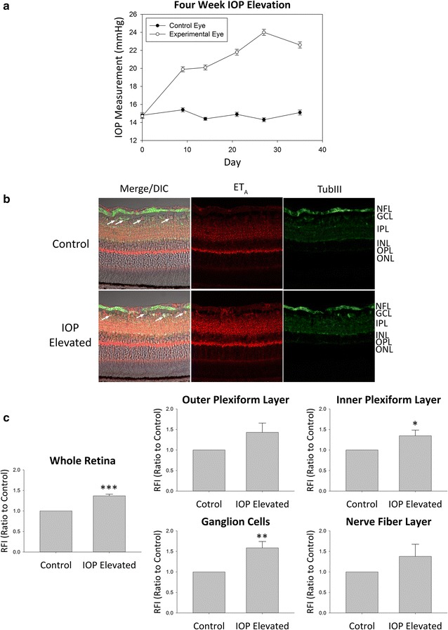
Fig. 2.

ETA expression in retinas of adult Brown Norway rats following 4 week IOP elevation. a Representative graph of IOP measurements for IOP elevated (white circles) and contralateral control (black circles) eyes in adult male Brown Norway rats. b Representative images. Immunostaining of retina sections probed for ETA receptors (red fluorescence) and β-III-tubulin (green fluorescence) following 4 weeks of IOP elevation. Arrows point to RGCs. c Relative fluorescent intensity for the retina, OPL, IPL, GCs and NFL. Bars represent mean ± SEM (n = 4 animals/group). Asterisks indicate statistical significance *p < 0.05; **p < 0.01; ***p < 0.001 by student’s t-test. ONL outer nuclear layer, OPL outer plexiform layer, INL inner nuclear layer, IPL inner plexiform layer, GCL ganglion cell layer, NFL nerve fiber layer, TubIII β-III-tubulin
