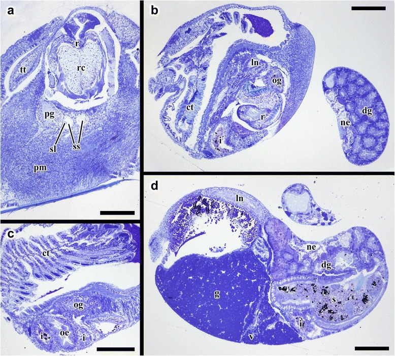
Fig. 7.

Transverse semi-thin sections from Gigantopelta chessoia. a. Posterior part of the head showing radula apparatus, pedal ganglion, and statocysts. b. Mid-body section showing the start of oesophageal gland. c. Mid-body section showing the oesophageal gland, sections through the digestive tract, and ctenidium. d. Section through the anterior part of the visceral sac showing the gonad, the ventricle and the digestive glands. Abbreviations: ct, ctenidium; dg, digestive gland; g, gonad; i, intestine; ln, lateral nerve cord; ne, nephridium; oe, oesophagus; og, oesophageal gland; pg, pedal ganglion; pm, pedal musculature; r, radula; rc, radula cartilage; re, rectum; s, stomach; sl, statolith; ss, statocyst; tt, cephalic tentacle; v, ventricle. Scale bars all 200 μm
