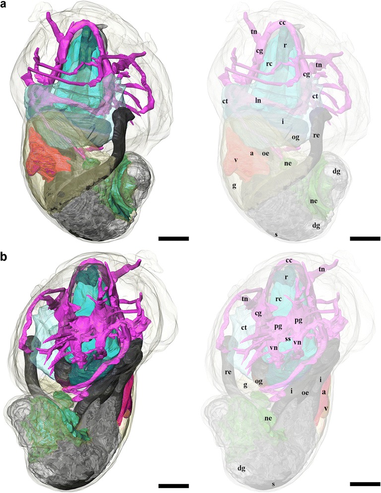
Fig. 8.

3D tomographic reconstruction of Gigantopelta chessoia, the full anatomical model in various views. Soft body outline (mantle and foot) shown in transparency for context. Ctenidium, anterior oesophagus, and digestive gland are rendered semi-transparent to show the structures underneath. For all parts, the tomographic model is shown to left and a second copy of the same view with labelled parts shown to right. a. Dorsal view; b. Ventral view. Colour groups correspond to specific anatomical systems: grey/black, digestive tract; brown, oesophageal gland; translucent blue, ctenidium; red, heart; yellow, gonad; green, nephridium; fuchsia/blue, nervous and sensory systems. Abbreviations: a, auricle; cc, cerebral commissure; cg, cephalic ganglion; ct, ctenidium; dg, digestive gland; g, gonad; i, intestine; ln, lateral nerve cord; ne, nephridium; oe, oesophagus; og, oesophageal gland; pg, pedal ganglion; r, radula; rc, radula cartilage; re, rectum; s, stomach; si, blood sinus; ss, statocyst; tn, tentacular nerves; v, ventricle; vn, ventral nerve cord. Scale bars, all 250 μm
