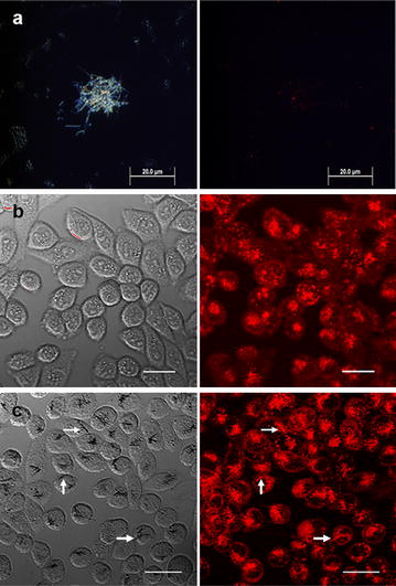
Fig. 5.

Confocal images of SiNWs after incubation with CHO-β cells. a Dark field (left) and fluorescence (right) images of the SiNW-NH2 after 1 h of incubation with DiI at 37 °C. b The transmission and fluorescence images of the control and c after 10 h of incubation with SiNW-NH2 at 37 °C. Arrows represent location of wires. Scale bars 20 µm
