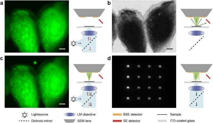
Figure 1. Working principle of cathodoluminescence for automated alignment.
(a) Fluorescence microscopy of MDCK cells expressing paxilin-GFP performed in-situ in a SEM. (b) The same area visualized with SEM. (c) Fluorescence microscopy as in (a) but with simultaneously the electron beam in spot mode. Due to substrate cathodoluminescence (CL), the position of the electron beam can be detected in the optical detection channel. (d) By quickly alternating the position of the electron beam to different locations in spot mode, multiple CL pointers can be detected simultaneously in a single acquisition using the light microscope. SE, secondary electrons; BSE, back-scattered electrons; ITO, indium tin oxide. Scale bars are 10 μm.

