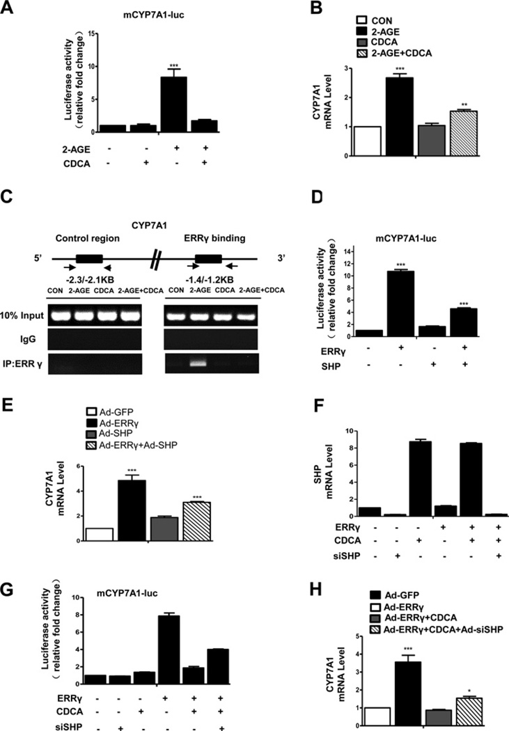
Figure 4. ERRγ-mediated induction of CYP7A1 gene expression is inhibited by SHP.
(A) AML12 cells were transfected with mCYP7A1-luc (−3.2 kb to +234 bp). At 36 h post transfection, cells were treated with 2-AGE (10 µM) in the presence or absence of CDCA (25 µM). (B) AML12 cells were treated with 2-AGE (10 µM) in the presence or absence of CDCA (25 µM) and CYP7A1 expression was analysed by qPCR. **P < 0.01; ***P < 0.001. (C) ChIP assay. AML12 cells were treated with 2-AGE (10 µM) in the presence or absence of CDCA (25 µM). Input represents 10% of purified DNA in each sample. Cell extracts were immunoprecipitated with anti-ERRγ antibody and purified DNA samples were employed for PCR with primers binding to ERRE1 (−1.4 kb to −1.2 kb) and distal site (−2.3 kb to −2.1 kb) on the CYP7A1 gene promoter. (D) AML12 cells were transfected with mCYP7A1-luc (−3.2 kb to +234 bp) and co-transfected with ERRγ or SHP expression vectors. (E) AML12 cells were infected with indicated adenoviruses and CYP7A1 expression was analysed by qPCR analysis. (F and G) AML12 cells were transfected with the si-SHP expression vector and after 24 h, the cells were co-transfected with mCYP7A1-luc (−3.2 kb to +234 bp) and ERRγ expression vector in the presence or absence of CDCA (25 µM). SHP mRNA level analysed by qPCR analysis. (H) AML12 cells were infected with Ad-ERRγ and Ad-shSHP in the presence or absence of CDCA (25 µM). CYP7A1 expression was analysed by qPCR analysis. *P < 0.05; ***P < 0.001. The cell lysates in (A, D and G) were utilized for luciferase and β-galactosidase assays.

