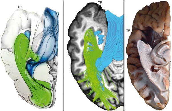
Figure 5.

Single subject (S5) MAGNET reconstruction of Meyer's loop shows close agreement with histological studies. Left: Anatomical drawing courtesy of Dr. Patrick Roth. Middle: Streamlines generated using MAGNET. Ventricle segmentation (blue) was added to display the relationship between Meyer's loop and the inferior horn. Colormap: T1‐weighted image. Right: Klingler dissection (ex vivo human brain, inferior view) of the optic radiation reveals a large and angulated anterior extent of Meyer's loop. Adapted from: Goga and Ture [2015], “The anatomy of Meyer's loop revisited: changing the anatomical paradigm of the temporal loop based on evidence from fiber microdissection,” J Neurosurg 2015;122:1253–1262 with permission of Rockwater, Inc. Note that the anatomical drawing omits the direct pathway linking the LGN to the occipital cortex. [Color figure can be viewed at http://wileyonlinelibrary.com]
