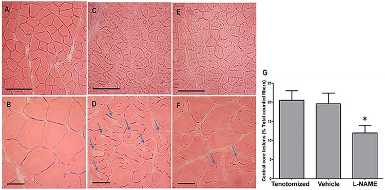
Figure 4. Analysis of central core lesion (CCL) occurrence on day 14 after injury. Control group (A, B), tenotomized group (C, D) and L-NAME group (E, F). Muscle fibers from the L-NAME group displayed remarkable histological alterations characteristic of CCL. Although CCLs were present in the L-NAME group, the occurrence of lesions was smaller than in the control group. Arrows: CCL in muscle fiber. Scale bar: 200 μm (A, C, E). Scale bar: 50 μm (B, D, F). n≥4 rats/group. G, Quantification of muscle fibers with CCL. A total of 180 fibers were evaluated from rats that underwent tenotomy (tenotomized, vehicle and L-NAME groups). Tenotomized and vehicle groups showed about 20% of muscle fibers with CCL. Treatment with L-NAME displayed a significant reduction of fibers with CCL (about 12%). Data are reported as means±SD. *P<0.05 vs tenotomized and vehicle (ANOVA-Bonferroni).