Fig. 1.
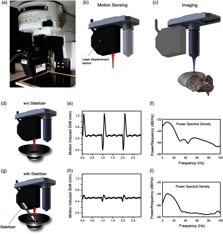
Axial motion characterization system for in vivo imaging. (a) The setup is fully integrated within a commercial confocal/two photon microscope (FV1000, Olympus). A laser displacement sensor is mounted onto a dual slide nosepiece, collinear with the imaging objective. (b) During motion characterization, the sensor is positioned over the imaging organ, directly measuring the total displacement along the axial direction. (c) After mouse or stabilizer readjustment to reduce axial motion components and to improve reproducibility over the physiological cycles, the imaging objective is slid in position in the same spot, where the sensor was focused and image acquisition is performed. (d–i) For setup testing and proof-of-principle repetitive induced artificial motion was used. A sample fixed on top of a loudspeaker (d,g) is moved vertically with a synthetic waveform producing 1 and 5 Hz sinusoidal motion components, approximately equal to the mouse respiratory and cardiac frequencies. (e, h) Amplitude measurements and (f, i) frequency analysis are performed without (d) and with (g) the presence of a mechanical stabilizer.

