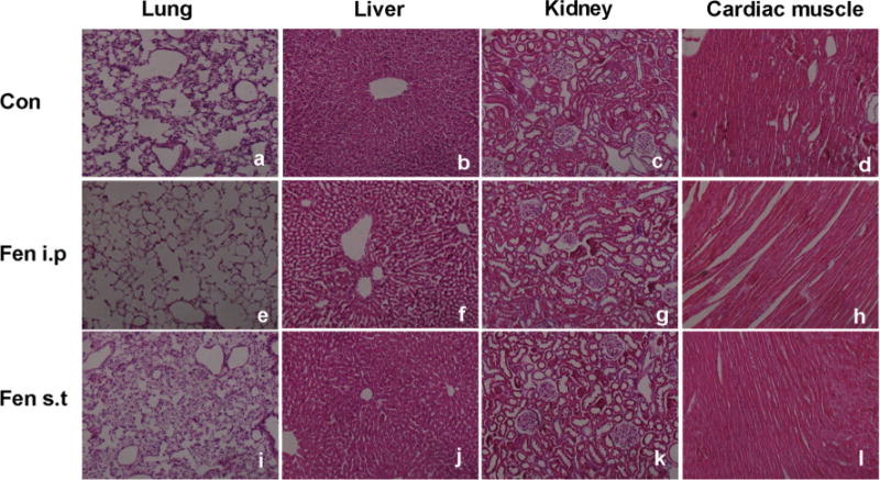
Fig. 9.

Pathological changes in peripheral organs of rats exposed to fenpropathrin by i.p or ST infusion, based on H&E staining. a–d Pathological morphology of the lung, liver, kidney, and cardiac muscle in the Con group. e–h Pathological morphology of the lung, liver, kidney, and cardiac muscle in fenpropathrin i.p group. i–l Pathological morphology of the lung, liver, kidney, and cardiac muscle in fenpropathrin ST group
