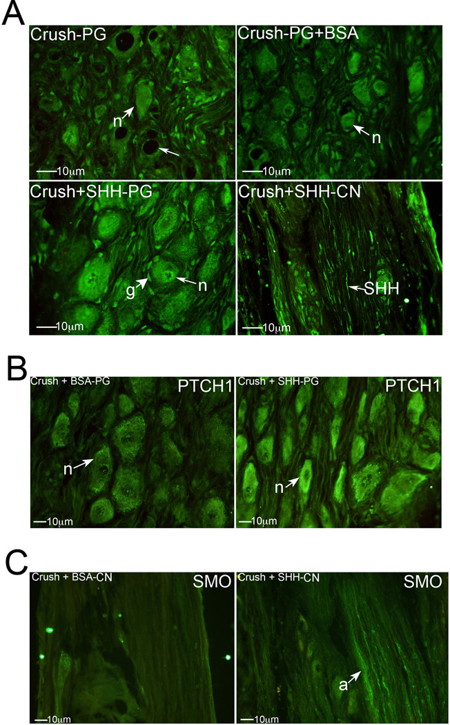
Figure 3.
(A) Immunohistochemical analysis of SHH protein in PG tissue of rats that under went CN crush (top left), CN crush with BSA protein treatment (control, top right), or CN crush with SHH protein treatment (bottom left), and CN tissue from rats that underwent CN crush with SHH protein treatment (bottom right). SHH protein was taken up at the crush site and underwent retrograde transport (bottom right) to PG neurons to maintain normal signaling (bottom left). (B) Immunohistochemical analysis of PTCH1 protein in PG tissue from rats that underwent CN crush with BSA treatment (control) or CN crush with SHH treatment, by PA (400× magnification). (C) Immunohistochemical analysis of SMO protein in CN tissue of rats that underwent CN crush with either BSA (control) or SHH protein treatment by PA. Anterograde transport of SMO stops with CN injury (left) and is maintained with SHH PA treatment (right) of the CN (400× magnification). Arrows indicate staining. n=neuron. g=glial cell.

