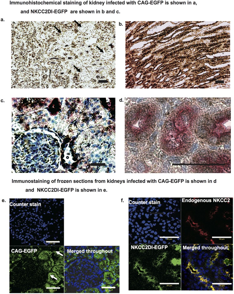
Fig 3. Immunohistochemistry of the kidneys.
a. Paraffin-embedded sections of the kidneys infected with CAG-EGFP adenovirus. The arrow indicates glomerulus. Adenovirus-derived EGFP was stained in brown. b and c. Paraffin embedded sections of the kidney infected with NKCC2DI-EGFP adenovirus. Adenovirus-derived EGFP is shown in brown and endogenous NKCC2 is shown in pink. e and f. Immunostaining of frozen kidney sections infected with CAG-EGFP is shown in e and NKCC2DI-EGFP is shown in f. e.Green indicates CAG promoter derived EGRP and f. Green indicates NKCC2 promoter-derived EGFP. f. Red indicates endogenous NKCC2. e and f. Blue indicates nucleus.

