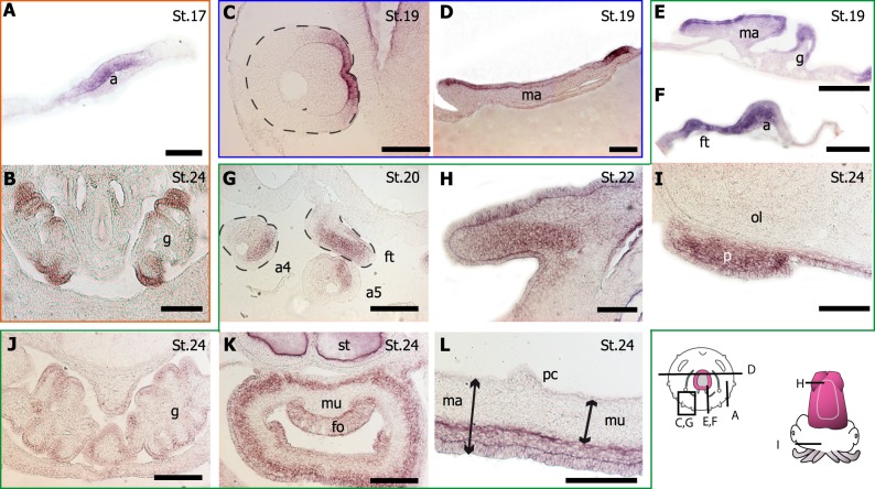
Fig 6. Pax gene expression in non neuronal structures.
(A, B) Pax6; (C, D) Pax3/7; (E-L) Pax2/5/8. Stages are indicated by St#. Note the artefact in statocyst (st) in K. Scale 150 μm, except E, F, G: 300 μm. Pax expression in arm (a): Pax6 (A. See also Fig 5C), Pax3/7 (C) and Pax2/5/8 (F, G a4 and a5. See also Fig 5I). Pax expression in mantle (ma): Pax3/7 (D) and Pax2/5/8 (E, L). Pax expression in gill (g): Pax6 (B) and Pax2/5/8 (E, J). Dynamic of Pax2/5/8 expression in funnel tube (ft): compare F, G and K (fo: funnel organ). Pax2/5/8 is also expressed in fin (H), arm pillars (p) (I). ol: optic lobe. mu: muscle. pc: pallial cavity. ss: shell sac.

