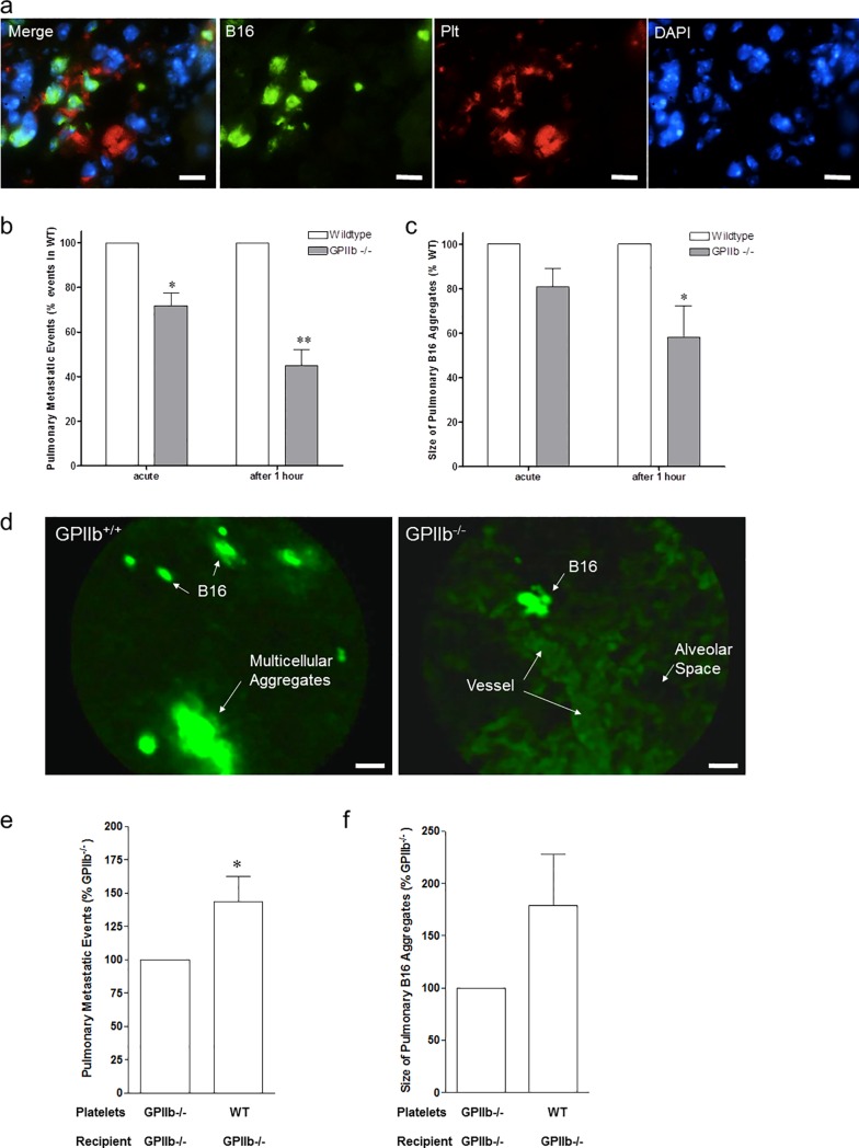
Fig 2. Platelet-tumor-aggregate formation in vivo.
a) GFP-transfected B16-D5 melanoma cells were injected intravenously into wildtype mice. After 1 hour, lung tissue was obtained for immunofluorescence analysis. Photomicrographs show mouse lung tissue stained with antibodies directed against platelet GPIIb (CD41, red) and B16-D5 (GFP, green); nuclei were stained with DAPI (blue). Bars, 40μm. Images were taken using a Leica DMRB epifluoresence microscope, 20x objective. b-d) Arrest of DCF-tagged B16-D5 melanoma cells was visualized in the pulmonary vasculature by intravital confocal videofluorescence microscopy (IVM) in GPIIb+/+ and GPIIb-/- littermate mice immediately and after 1 hour. b) Number of metastatic events was quantified. Results are given as percentage of firmly adherent B16-D5 in GPIIb-/- mice compared to its WT littermate (n = 5–6 experiments per group; *P<0.01 acute; **P<0.001 after 1 hour). c) Size of B16-aggregates was quantified. Results are given as percentage of B16-aggregate size in GPIIb-/- mice compared to its WT littermate (n = 5–6; *P<0.05 after 1 hour). d) Photomicrographs show representative IVM images obtained in GPIIb+/+ and GPIIb-/-. In GPIIb+/+ mice, arrest of large multicellular aggregates is frequently observed (left). Arrest of DCF-tagged B16-D5 is visualized in precapillary vessels (right). Bars, 20μm. d e-f) GPIIb+/+ (WT) or GPIIb-/- platelets were injected into GPIIb-/- mice just prior to administration of DCF-labeled B16-D5. IVM was performed immediately and after 1 hour. e) Number of metastatic events was quantified. Results are given as percentage of firmly adherent B16-D5 in GPIIb-/- littermates receiving GPIIb-/- platelets (n = 4; *P<0.05). f) Size of B16-aggregates was quantified. Results are given as percentage of B16-aggregate size in GPIIb-/- littermates receiving GPIIb-/- platelets (n = 3; P = n.s.).

