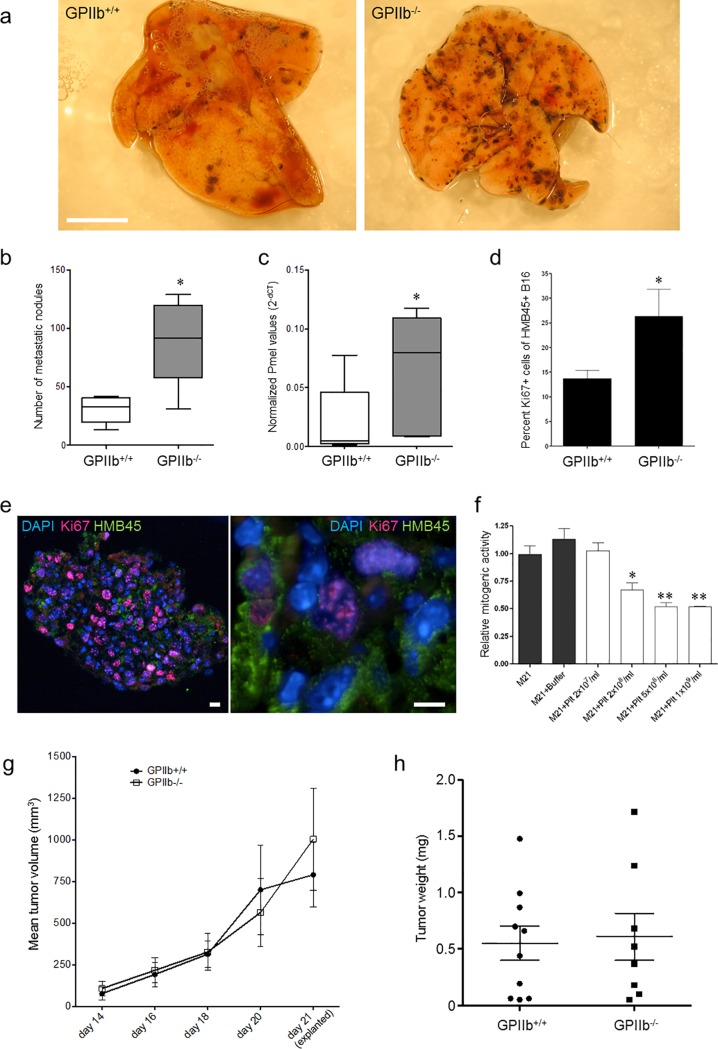
Fig 3. Effect of platelet GPIIb on melanoma metastasis formation.
a)-c) B16-D5 melanoma cells were injected intravenously into GPIIb+/+ or GPIIb-/- littermate mice. After 10 days, metastasis formation was analyzed. a) Photomicrographs show representative lung images. Bars, 0.5 cm. b) Quantification of metastatic nodules in the lung (n = 5; *P<0.05). Boxes indicate median, whiskers indicate min and max values. c) Quantification of Pmel mRNA expression. Individual Pmel data normalized to beta-actin is indicated as 2(-ΔCt) (n = 5; *P<0.05). Boxes indicate median, whiskers indicate min and max values. d-e) Pulmonary melanoma proliferation was determined by analyzing Ki67 expression in HMB-45-positive melanoma cells 10 days after intravenous seeding (n = 5; *P<0.05). Photomicrographs show fluorescence microscopy images of mouse lung tissue stained with antibodies directed against melanoma HMB-45 (green) and Ki67 (red); nuclei were stained with DAPI (blue). Images show 3μm optical sections. Left image, overview of lung tissue section; right image, magnification. Bars, 100μm. Images were taken using a Leica DMRB epifluoresence microscope, 20x and 40x objective. f) Effect of platelets on M21 mitogenesis in vitro. Serum-starved M21 cells were grown in the absence or presence of increasing concentrations of washed human platelet and BrdU uptake was quantified (*P<0.05, **P<0.01). g-h) Effect of platelet GPIIb on subcutaneous melanoma growth. g) B16-D5 cells were injected subcutaneously in the right flank of wildtype and GPIIb-/- littermate mice and tumor growth surveyed using a digital scaliper. h) 21 days after tumor cell seeding tissues were explanted. Tumor weight was measured indicating no difference in subcutaneous tumor growth (P = n.s.).

