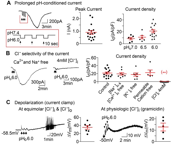
Figure 1. A prolonged pHo-conditioned inward current and depolarizations.
(A) A representative inward current evoked from a nodose neuron after 3 brief sequential exposures to extracellular pHo 6.0, indicated by the vertical black arrows. The membrane potential was held at –60 mV. The peak currents seen in 17 of 22 individual neurons are shown. The current density is pHo dependent and averaged 8.5 ± 2.1 pA/pF for pHo 7.0 (n = 2 mice); 10.8 ± 2.9 pA/pF for pHo 6.5 (n = 2 mice); 21.2 ± 2.7 pA/pF for pHo 6.0 (n = 6 mice, P = 0.037 by ANOVA). (B) The currents evoked in control solutions (21.2 ± 2.7 pA/pF, n = 3 mice) are maintained in extracellular Ca2+ and Na+ free solution (19.3 ± 3.1 pA/pF, n = 3 mice), in intracellular K+ free solutions (17.4 ± 4.2 pA/pF, n = 5 mice ), and in solutions free of all permeable cations (25.9 ± 6.6 pA/pF, n = 3 mice ) but are absent in solutions of 4 mM intracellular Cl– (0.6 ± 0.2 pA/pF, n = 5 mice, **P < 0.001 vs. control by ANOVA). (C) Under current clamp conditions, progressive depolarizations and action potentials were evoked following the transient sequential pHo 6.0 applications (black arrows). The tracing and panel to the left show the mean maximum depolarizations of 35.2 ± 4.4 mV (n = 5 mice) at equimolar (133 mM) [Cl–]i and [Cl–]o and an equilibrium potential of ~0 mV. The tracing and panel to the right show the membrane potential and maximum depolarizations recorded with the perforated patch clamp using gramicidin to maintain physiologic intracellular [Cl–]i and an equilibrium potential that is more negative hence a lesser maximal depolarization (12.7 ± 3.4 mV, n = 2 mice). All panels include responses of individual neurons and the means ± SE.

