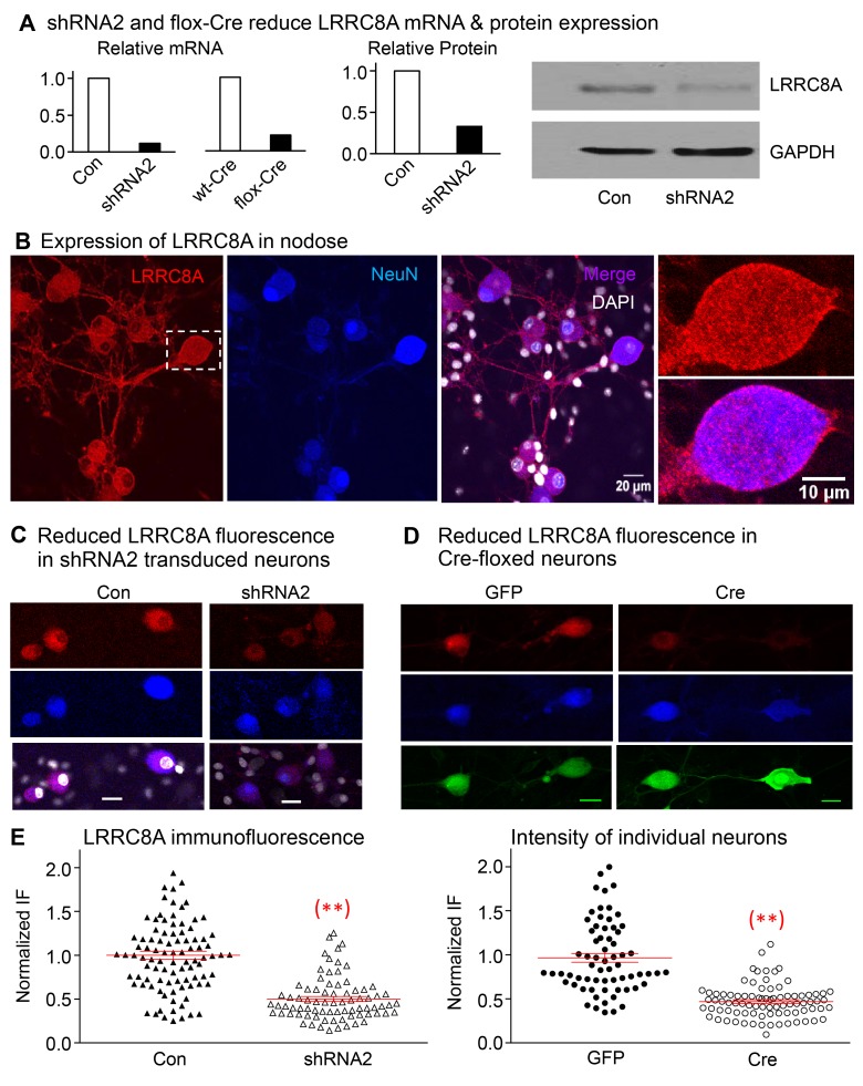
Figure 4. Expression of LRRC8A is significantly reduced in neurons transfected with shRNA2 and in Cre-floxed neurons.
(A) The relative mRNA of LRRC8A is reduced from 100% in control to 12% in shRNA2-transduced neurons and from 100% in WT-Cre to 22% in flox-Cre neurons targeting the shRNA2 sequence used (5 mice were used in each group). The relative protein is reduced from 100% in control to 33% in shRNA2-transduced neurons (5 mice in each group), and the Western blot result shown confirms the reduction in LRRC8A relative to GAPDH. (B) The immunofluorescence of LRRC8A (red) is expressed in the soma of nodose ganglia neurons and their neurites. The expression is colocalized with neuronal marker NeuN (blue) and is not apparent in nonneuronal cells (DAPI panel). The boxed neuron in the left panel is magnified in the 2 panels on the right to show the prominent membrane expression of LRRC8A (see also Supplemental Figure 4 for immunofluorescence of LRRC8A in membranes of HEK293 cells). (C) The red fluorescence of LRRC8A is shown in neurons transduced with control vector without shRNA (NeuN in blue, left panels). The signal of LRRC8A is reduced in neurons transduced with shRNA2 (right panels) (horizontal bars in lower panels represent 20 μm). (D) Nodose neurons from floxed mice show clear staining of LRRC8A (red) in GFP-transduced group (left panels) and reduced staining in the Cre-transduced group (right panels). Red fluorescence of LRRC8A colocalizes with the neuronal marker (blue) NeuN. (E) The normalized fluorescence intensity of LRRC8A in control neurons (Con) is reduced significantly from 1.00 ± 0.06 (n = 67 neurons from 3 mice) to 0.47 ± 0.02 (n = 84 neurons from 3 mice, **P < 0.001) in Cre-transduced neurons, and from 1.00 ± 0.05 (n = 94 from 2 mice) to 0.50 ± 0.03 (n = 78, **P < 0.001 from 2 mice) in neurons transduced with shRNA2.

