Fig. 4.
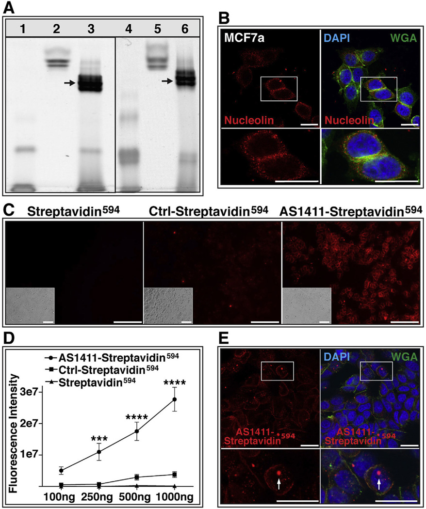
AS1411 delivers protein to cells in vitro. (A) Native polyacrylamide gel confirms conjugation of streptavidin594 with biotinylated AS1411. Lane 1: biotinylated AS1411; Lanes 2 and 5: Streptavidin594; Lane 3: AS1411-Streptavidin594 conjugate; Lane 4: biotinylated control oligonucleotide; Lane 6: Control-Streptavidin594 conjugate;. Arrows indicate conjugation products. (B) MCF7a cells probed with antibody against nucleolin (red) or stained with wheat germ agglutinin (WGA; green) and DAPI (blue). Scale bar = 20 µm (C) MCF7a cells incubated for 1 h with 1000 ng of Streptavidin594, Ctrl-Streptavidin594, or AS1411-Streptavidin594. Bright-field images shown in insert Scale bar = 120 µm. (D) Quantification of raw fluorescence intensity of MCF7a cells incubated with increasing doses of Streptavidin594, Ctrl-Streptavidin594, or AS1411-Streptavidin594 (study performed twice in triplicate) (*** = p < 0.001; **** = p < 0.0001). (E) Confocal microscopy of AS1411-Streptavidin594 (red) incubated cells stained with WGA (green) and DAPI (blue). Arrow indicates nuclear localization of AS1411-Streptavidin594. Scale bar = 20 µm. Ctrl-Control. (For interpretation of the references to color in this figure legend, the reader is referred to the web version of this article.)

