Fig. 7.
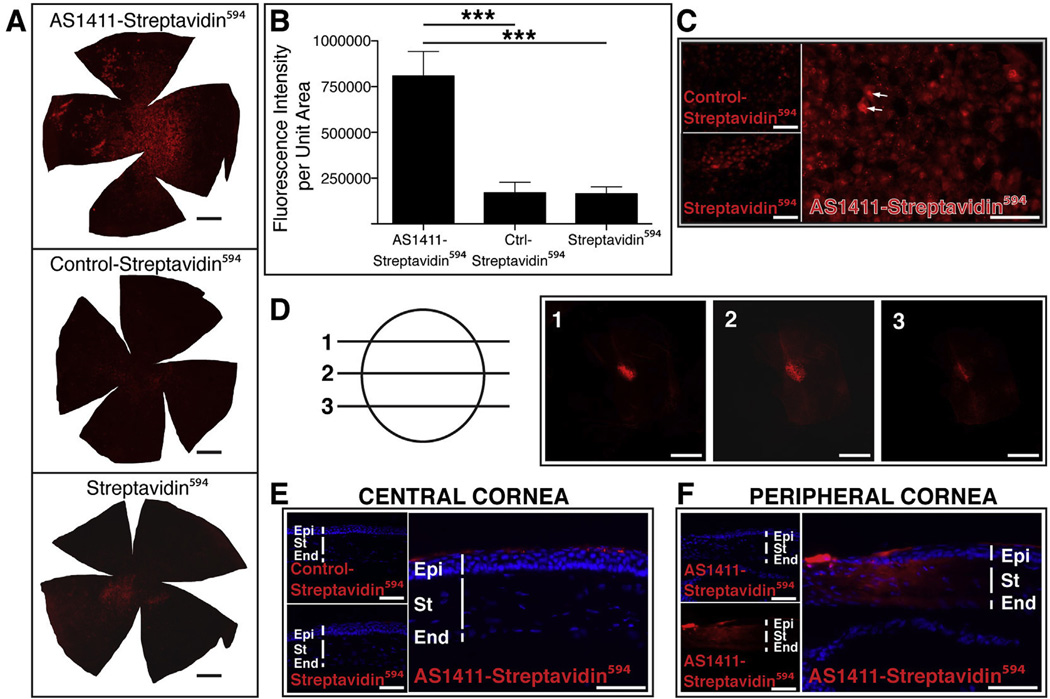
Topical Application of AS1411-streptavidin594 results in uptake by corneal cells and nuclear localization in vivo. (A) Corneal flat mounts harvested 2 h post topical application of 5 µg of AS1411-Streptavidin594, Control-Streptavidin594 or Streptavidin594. Scale bar = 0.5 mm. (B) Quantitation of the intensity of fluorescence of streptavidin594 conjugates per area of corneal flatmount (n = 6 per condition, *** = p < 0.001). (C) Higher magnification images of AS1411-Streptavidin594, Control-Streptavidin594 or Streptavidin594 treated corneal flat mounts. Scale bar =120 µm. Arrows indicate examples of nuclear binding. (D) Confocal series of a flatmount of AS1411-Streptavidin594 treated cornea indicating nuclear localization within a corneal epithelial cell. Scale bar = 20 µm. (E) Transverse sections of corneas treated topically with AS1411-Streptavidin594, Control-Streptavidin594 or Streptavidin594 (n = 4 eyes/2 mice per condition). Scale bar = 60 µm. (F) Transverse section of AS1411-strepatividin594 treated cornea showing transduction of stroma and endothelium at the corneal limbus. Scale bar = 60 µm. Ctrl-Control; Epi-epithelium; St-stroma; End-endothelium.

