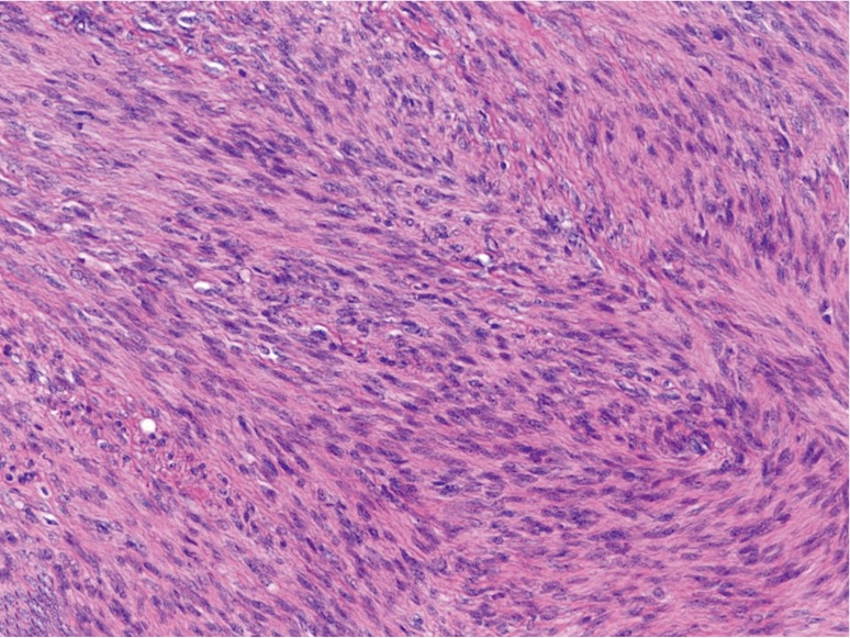
Figure 1.

Hepatic lesion showing interlacing fascicles of spindle cells with eosinophilic cytoplasm and elongated, blunt-ended nuclei with stippled chromatin. There are rare interspersed blood vessels within the fascicles, and mitotic figures are inconspicuous (H&E, 200×).
