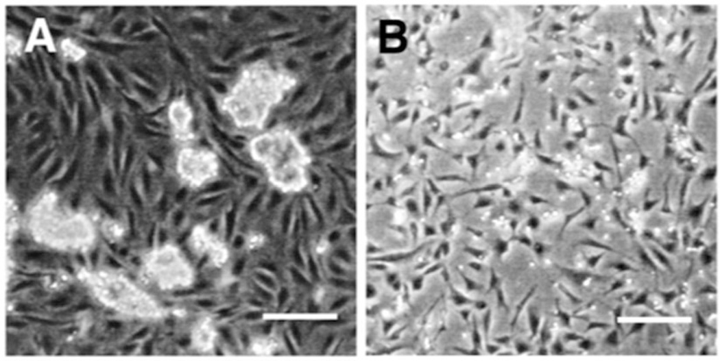
Fig. 1.
hESC coculture and CD271+ cells after isolation. (a) hESC coculture on PA6 cell monolayer. hESC retain colony-like morphology while the underlying PA6 cells remain confluent and spindle-like. (b) CD271+ Cells grown in serum free medium after isolation via MACS cell separator. Scale bars are 200 µm. Figure from ref. (6)

