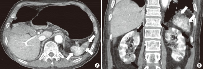
Fig. 1.
MDCT images in 85-year-old man with an atrial fibrillation. Axial image (A) shows multiple infarctions in the spleen (white arrows). Coronal image (B) shows multifocal wedge-shaped infarctions in bilateral kidneys (black arrows) and multiple infarctions in the spleen (white arrows). Renal infarction caused by an embolism was diagnosed.
MDCT = multidetector computed tomography.

