Fig. 1.

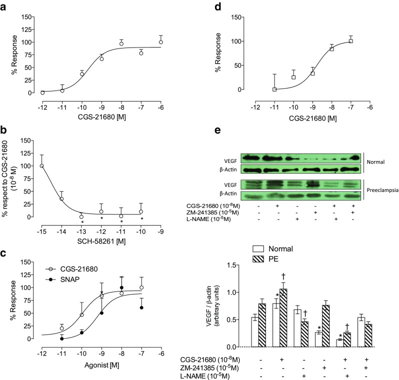
A2A-NO-VEGF pathway in feto-placental endothelial cells. Endothelial cells from human umbilical vein (HUVEC) were isolated from normal pregnancies and used for cell proliferation analysis by incorporation of bromouridine. Cells were incubated for 24 h with a CGS-21680 (10−6 to 10−12 M) and b co-incubated with CGS-21680 (10−8 M, maximum dose) and SCH-58261 in dose response (10−15 to 10−10 M) or c CGS-21680 (10−7 to 10−12 M, white circles) or SNAP (10−7 to 10−11 M, black circles). d Placental microvascular endothelial cells (hPMEC) taken from normal pregnancies (white bar) and pre-eclamptic (hatched bar) were extracted as described in Methods section. Cell proliferation quantified by incorporating bromouridine in the presence of CGS-21896 (10−6 to 10−11 M). e Representative image of western blot analysis for VEGF and its respective load control β-actin in the presence of CGS-21680 (10−8 M) and/or ZM-241385 (10−6 M) and/or L-NAME (10−5 M) in cells from normal and pre-eclamptic pregnancies (top panel). In the lower panel, densitometry of VEGF/β-actin ratio treated as indicated in the top panel. The presence of drugs is marked with (+) and absence with (−). Results are expressed as medians and interquartile ranges. In b, *p < 0.05 vs 100 % response. n = 5 for each assay in duplicate. In d, n = 4. In e, n = 3 per group in duplicated
