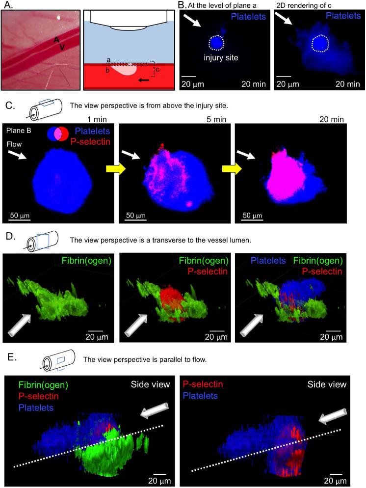
Figure 1. Laser injury of the mouse femoral artery.
(A) A representative image of the mouse femoral artery (A) and vein (V), and schematic of microscope orientation to the vessel and injury site, and the various focal planes used for imaging (A-C). Note that the site of injury and, therefore, the thrombus core are closer to the microscope objective than the thrombus shell. (B) Representative images of the resulting thrombus (blue) 20 minutes post-injury. A confocal slice at the vessel wall (plane a, left) shows the injury site area, and the compressed 3D stack of the thrombus (plane c, right) shows the full size of the resulting thrombus. Plane b is at the inner surface of the vessel wall. (C) A time course of representative images showing platelet accumulation (blue) and P-selectin exposure (red) following laser injury. (D) A 3D rendering of a representative thrombus showing fibrin(ogen) (green), P-selectin (red), and platelets (blue) 20 minutes post-injury from the front, and (E) from the side.

