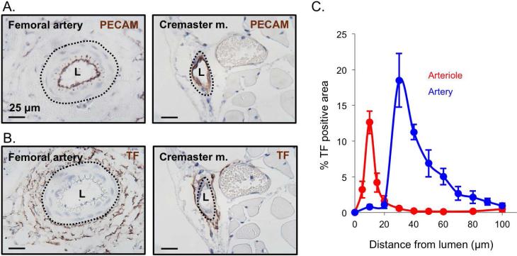
Figure 4. Tissue factor distribution in the macro- and microcirculation.
Representative images of mouse femoral artery and cremaster arteriole showing (A) PECAM and (B) tissue factor staining of vessel endothelium (brown) and vessel lumen (L). (C) Quantification of the percent positive area of tissue factor radiating out from the vessel lumen center for the femoral artery (blue) and cremaster arterioles (red) (+/− SEM, n = 5 femoral and 5 cremaster).

