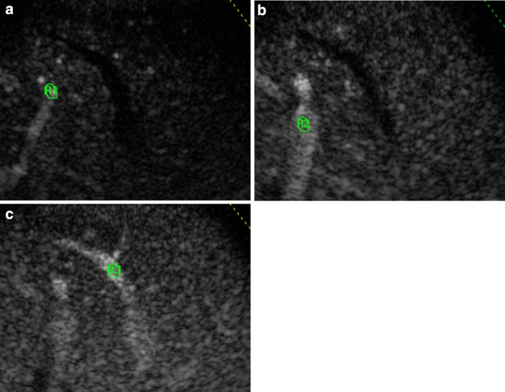
Fig. 1.
Regions of interest (ROI) are traced on a branch of the hepatic artery (a), portal (b) and hepatic vein (c), simultaneously scanned using an intercostal section. A region alignment to the three vessels was obtained for all frames of the recorded clip: frame by frame, ROIs were positioned perfectly inside the vessels, to minimize the effect due to parenchymal intensity

