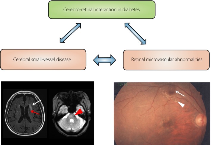
Figure 6.

Cerebro–retinal interaction in diabetes. A 76‐year‐old woman with simple diabetic retinopathy. (a) Magnetic resonance imaging expressions of cerebral small vessel disease including silent brain infarction (red arrow), white matter lesion (white arrow) and microbleed (arrow head). (b) Retinal photograph of diabetic retinopathy signs showing microaneurysm and retinal hemorrhages (arrow), and hard exudates (arrow head).
