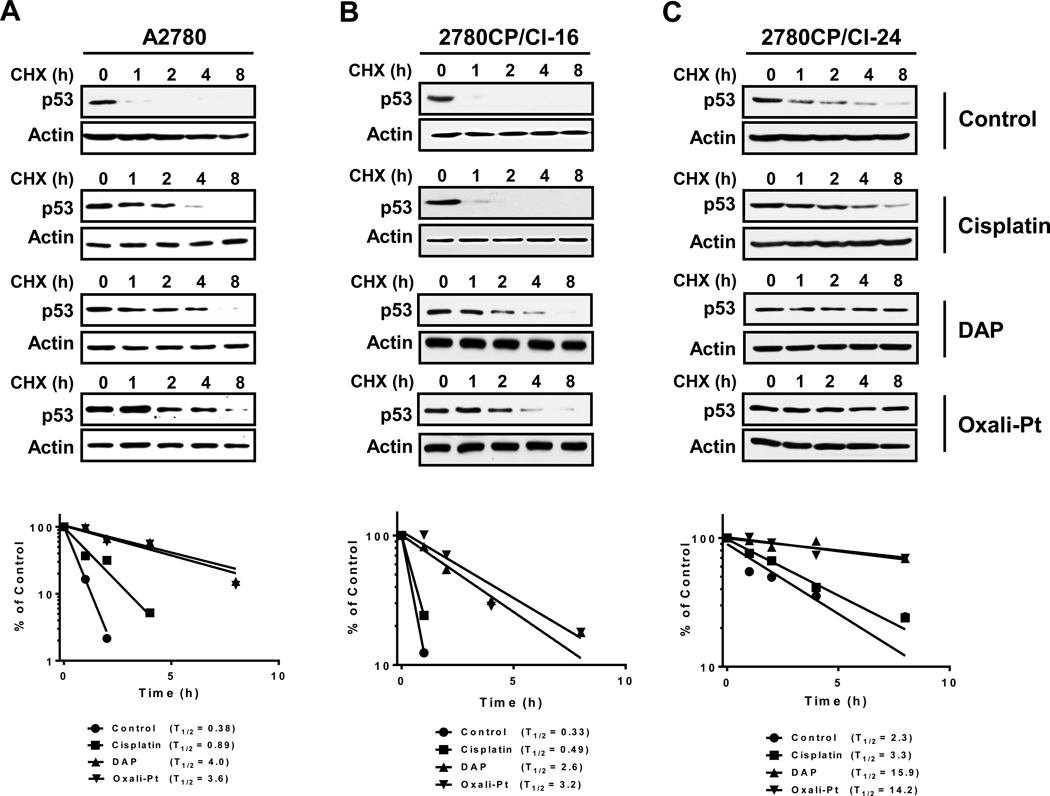
Figure 3.
Determination of p53 half-life in tumor cell lines exposed to platinum drugs. A2780 (A), 2780CP/Cl-16 (B) and 2780CP/Cl-24 (C) cells treated with cisplatin, DAP or oxaliplatin for 24 hours were additionally exposed to 4 µM cycloheximide (CHX) and then harvested at the indicated times. Cell lysates were prepared and resolved by SDS-PAGE, and p53 levels examined by immunoreaction. The p53 and β-actin signals in the immunoblots were quantified by densitometry, the ratios of p53/β-actin were normalized to time zero and plotted against time.

