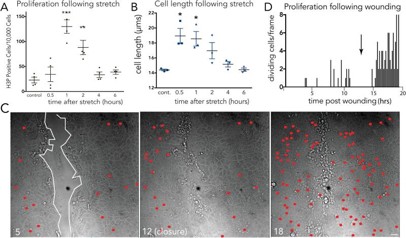
Figure 1. Mechanical stretch induces epithelial monolayers to rapidly divide.
(A) Proliferation rates (A) and cell lengths (B) at various times following stretch show that stretch-induced cell divisions return cell densities to control levels, where values are the averages of 3 experiments measuring the mean of 6 areas, error bars = s.e.m. P-values from unpaired T-tests compared to control are ***<0.0005, **<0.005, *<0.01. (C) Stills showing cumulatively where and when cells divide (red dots) during wound healing of an MDCK monolayer, where wound edge (highlighted with white line) with time in hours. (D) Graph (one of 14 similar) of cell divisions after monolayer wounding, with arrow indicating wound closure.

