Fig. 5.
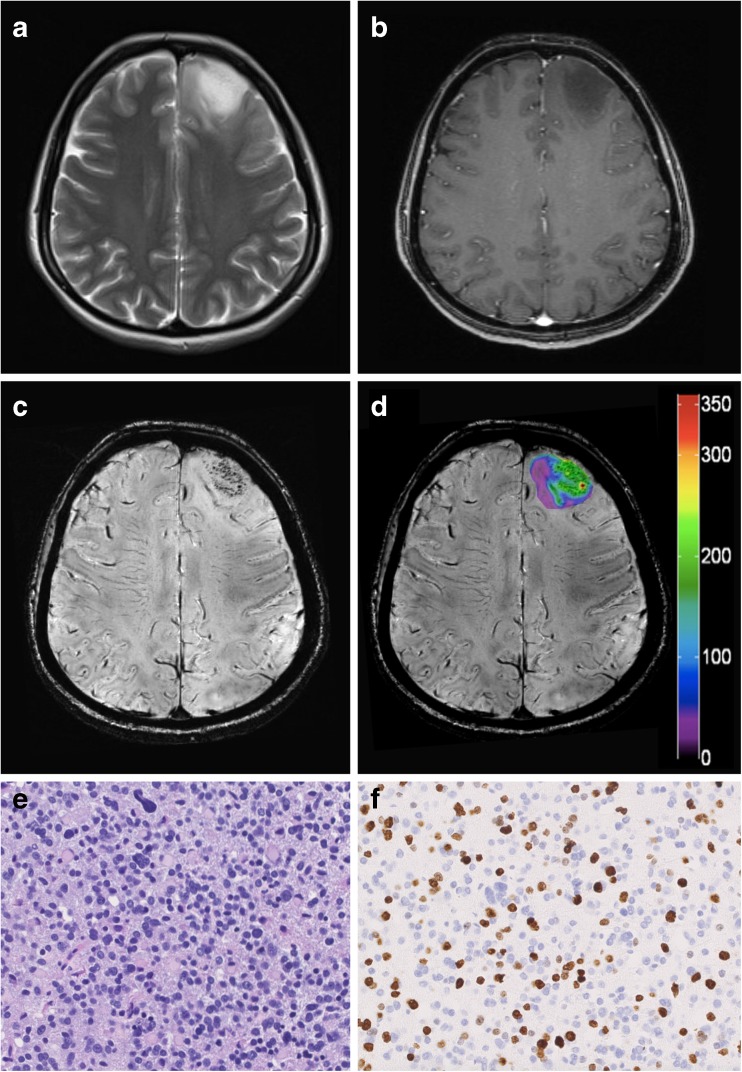
Preoperative identification of early malignant transformation in an initially suspected LGG on conventional MRI. (A) Preoperative axial T2-weighted MR images and (B) contrast-enhanced T1-weighted sequences reveal a left frontal hyperintense lesion with non-significant (none) CE. (C) Although no CE is visible on conventional MRI, 7 Tesla SWI depicts markedly increased intratumoural hypointense structures and (D) the corresponding colour-coded SWI-LIV map shows high values in the tumour ROI (mean SWI-LIV: 82.0). (E) Histopathological examination depicts already a HGG (anaplastic oligodendroglioma WHO grade III) with a (F) markedly increased proliferation rate (MIB-1 labelling index: 30 %). Original magnification of the H&E stain (E) and anti–Ki 67 stain (F) is × 200

