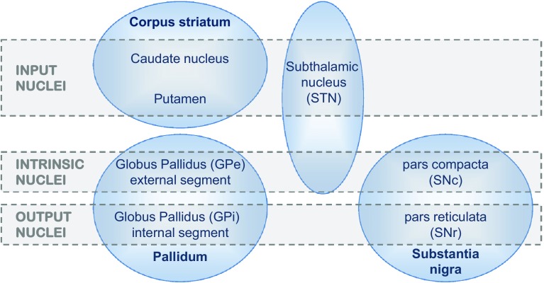
Fig. 1.
Components of the basal ganglia. The diagram distinguishes the anatomical identity of nuclei (shown in blue ovals) from their functional role, as assessed by input/output connectivity (indicated by grey bands). The corpus striatum can be considered a single nucleus, perforated by the internal capsule, and named for the strands of grey matter that stretch between the caudate and putamen. The caudate is typically referred to as a ‘nucleus’ whilst the putamen is not, though their cellular constitution is much the same. These two subdivisions are also known collectively as the dorsal striatum, as opposed to the ventral striatum which incorporates the nucleus accumbens (not shown here). Similarly, the two components of the output module, the substantia nigra pars reticulata (SNr), and the internal segment of the globus pallidus (GPi) also share a similar cellular composition and a continuous connectional topography, despite being quite separate anatomically. The subthalamic nucleus (STN) combines both extrinsic (cortical) and intrinsic inputs—the latter originating from another intrinsic nucleus, the external segment of the globus pallidus (GPe). Finally, the substantia nigra pars compacta (SNc) has reciprocal connections with the striatum, though it also receives extrinsic inputs. The striatum, GPe, GPi and SNr all comprise GABAergic projection neurons; the striatum also has several types of identified interneurons, one cholinergic plus three GABAergic. The STN is the only glutamatergic nucleus, comprising just one cell type. The SNc has dopaminergic projection neurons that issue collaterals to several BG nuclei in addition to their main target, the striatum

