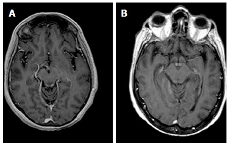
Figure 4.

Axial T1-weighted magnetic resonance images after contrast material administration in a patient with alcoholic Wernicke’s encephalopathy. A: Enhanced axial T1-weighted image shows contrast-enhancement of the periaqueductal grey matter; B: Enhanced axial T1-weighted image shows contrast-enhancement around the mammillary bodies.
