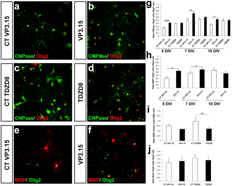
Figure 1. VP3.15 favors differentiation of murine OPCs without affecting their proliferation.
(a–d) Immunofluorescence images showing the expression of Olig2 and CNPase by differentiated OPCs from P0 mice after 5 DIV cultured in the presence of VP3.15 (a,b) or TDZD8 (c,d). (g) Quantification of CNPase+-Olig2+ cells with respect to the total number of Olig2+ cells at 5, 7 and 10 DIV. The presence of VP3.15 increases the number of oligodendrocytes in comparison with control conditions. TDZD8 did not induce changes in OPC differentiation. (e,f) Immunofluorescence images showing the expression of Olig2 and MBP by differentiated OPCs from P0 mice after 5DIV, cultured in the presence of VP3.15. (h) Quantification of MBP+-Olig2+ cells with respect to the total number of Olig2+ cells at 5, 7 and 10 DIV in the presence of VP3.15. (i) Determination of apoptotic oligodendroglia (Casp3+-A2B5+ cells). OPC survival was enhanced in the presence of TDZD8 only (1 μM). (j) Quantification of BrdU incorporation by double immunocytochemistry on OPCs from P0 mice. The presence of VP3.15 or TDZD8 did not modify the number of BrdU+-Olig2+ cells compared with their respective controls. Scale bar represents 25 μm for (a–f ). Values are given as mean ± SEM and the results of Student’s t-test are represented as *p < 0.05, **p < 0.01, and ***p < 0.001.

