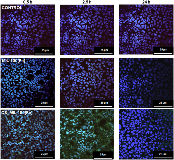
Figure 5. Confocal microscopy images of Caco-2 cells containing uncoated and CS-coated MIL-100(Fe) NPs observed by iron self-reflection signal (green channel) and the nucleus stained by DAPI (blue channel).
The images have been taken at different times: 0.5, 2.5 and 24 h. Moreover, the controls were obtained with cells (control) after 24 h. In all the cases, the scale bar corresponded to 25 μm. All the images were taken at 63X.

